| Nov 24, 2021 |
|
|
|
(Nanowerk Information) Most human illnesses may be traced to malfunctioning elements of a cell — a tumor is ready to develop as a result of a gene wasn’t precisely translated into a specific protein or a metabolic illness arises as a result of mitochondria aren’t firing correctly, for instance. However to know what elements of a cell can go incorrect in a illness, scientists first have to have a whole listing of elements.
|
|
By combining microscopy, biochemistry methods and synthetic intelligence, researchers at College of California San Diego College of Drugs and collaborators have taken what they suppose could turn into a big leap ahead within the understanding of human cells.
|
|
The approach, referred to as Multi-Scale Built-in Cell (MuSIC), is decsribed in (“A multi-scale map of cell construction fusing protein photos and interactions”).
|
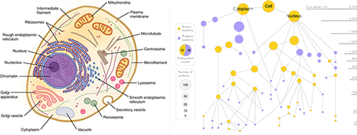 |
| Left: Basic textbook cell diagrams suggest all elements are clearly seen and outlined. (Credit score: OpenStax/Wikimedia). Proper: A brand new cell map generated by MuSIC technic reveals many novel elements. Gold nodes signify identified cell elements, purple nodes signify new elements. The scale of node displays variety of distinct proteins in that part. (Picture: UC San Diego Well being Sciences) (click on on picture to enlarge)
|
|
“In case you think about a cell, you in all probability image the colourful diagram in your cell biology textbook, with mitochondria, endoplasmic reticulum and nucleus. However is that the entire story? Positively not,” mentioned Trey Ideker, PhD, professor at UC San Diego College of Drugs and Moores Most cancers Heart. “Scientists have lengthy realized there’s extra that we don’t know than we all know, however now we lastly have a method to look deeper.”
|
|
Ideker led the examine with Emma Lundberg, PhD, of KTH Royal Institute of Expertise in Stockholm, Sweden and Stanford College.
|
|
Within the pilot examine, MuSIC revealed roughly 70 elements contained inside a human kidney cell line, half of which had by no means been seen earlier than. In a single instance, the researchers noticed a bunch of proteins forming an unfamiliar construction. Working with UC San Diego colleague Gene Yeo, PhD, they finally decided the construction to be a brand new advanced of proteins that binds RNA. The advanced is probably going concerned in splicing, an necessary mobile occasion that allows the interpretation of genes to proteins, and helps decide which genes are activated at which instances.
|
|
The insides of cells — and the numerous proteins discovered there — are usually studied utilizing one in every of two methods: microscope imaging or biophysical affiliation. With imaging, researchers add florescent tags of assorted colours to proteins of curiosity and observe their actions and associations throughout the microscope’s subject of view. To take a look at biophysical associations, researchers would possibly use an antibody particular to a protein to tug it out of the cell and see what else is hooked up to it.
|
|
The group has been concerned about mapping the internal workings of cells for a few years. What’s completely different about MuSIC is the usage of deep studying to map the cell straight from mobile microscopy photos.
|
|
“The mixture of those applied sciences is exclusive and highly effective as a result of it’s the primary time measurements at vastly completely different scales have been introduced collectively,” mentioned examine first writer Yue Qin, a Bioinformatics and Techniques Biology graduate scholar in Ideker’s lab.
|
|
Microscopes enable scientists to see right down to the extent of a single micron, in regards to the dimension of some organelles, comparable to mitochondria. Smaller components, comparable to particular person proteins and protein complexes, can’t be seen via a microscope. Biochemistry methods, which begin with a single protein, enable scientists to get right down to the nanometer scale. (A nanometer is one-billionth of a meter, or 1,000 microns.)
|
|
“However how do you bridge that hole from nanometer to micron scale? That has lengthy been an enormous hurdle within the organic sciences,” mentioned Ideker, who can also be founding father of the UC Most cancers Cell Map Initiative and the UC San Diego Heart for Computational Biology and Bioinformatics. “Seems you are able to do it with synthetic intelligence — information from a number of sources and asking the system to assemble it right into a mannequin of a cell.”
|
|
The group skilled the MuSIC synthetic intelligence platform to take a look at all the info and assemble a mannequin of the cell. The system doesn’t but map the cell contents to particular places, like a textbook diagram, partially as a result of their places aren’t essentially mounted. As a substitute, part places are fluid and alter relying on cell sort and scenario.
|
|
Ideker famous this was a pilot examine to check MuSIC. They’ve solely checked out 661 proteins and one cell sort.
|
|
“The clear subsequent step is to blow via all the human cell,” Ideker mentioned, “after which transfer to completely different cell sorts, folks and species. Finally we’d be capable to higher perceive the molecular foundation of many illnesses by evaluating what’s completely different between wholesome and diseased cells.”
|


